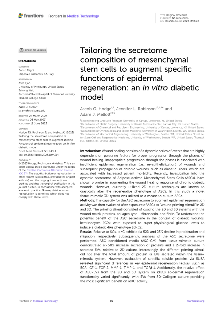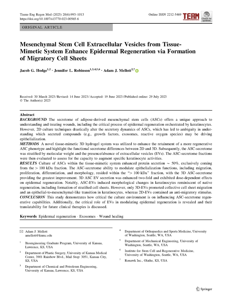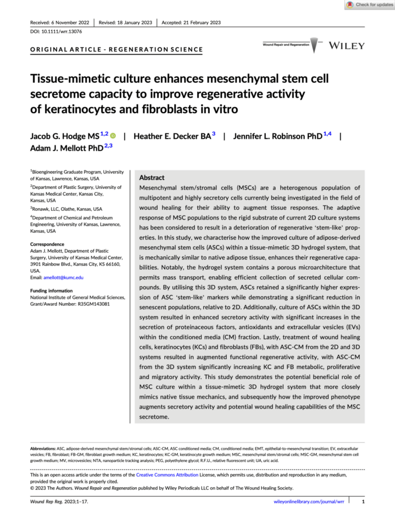
Jacob G. Hodge, Jennifer L. Robinson, Adam J. Mellott
Front. Med. Technol. 5:1194314. doi: 10.3389/fmedt.2023.1194314
RECEIVED 27 March 2023 | ACCEPTED 26 May 2023 | PUBLISHED 12 June 2023
Introduction & Background
Successful wound healing relies on the proper progression of wound healing phases where a diverse range of cells are correctly responding to signaling cues at the correct times. Unfortunately, some wound types and patient populations are susceptible to inadequate healing and chronic wound development. One such example is the development of diabetic ulcers, which are associated with increased patient morbidity.
A critical step in wound healing is the re-epithelialization of the wound site by keratinocytes, which restores the skin barrier and protects deeper tissue layers. For this process to occur successfully keratinocytes need to become more migratory and proliferative, so they can move to the wound site and increase in number. A secondary change is for keratinocytes to switch the specific subtypes of keratin they produce, so they are better able to re-epithelialize the wound. Signals from surrounding cell populations help to guide this process. Dysregulation of this signaling can lead to wounds that have protracted closure times or develop into non-healing wounds that do not close.
Diabetic wounds in particular have an improper imbalance of bioactive compounds (depleted growth factors, inflammatory cytokines, etc..), which can impair normal wound healing processes. High glucose levels are also known to have negative effects. These factors can result in improper wound healing processes – including altered keratinocyte differentiation and activity. Isolated keratinocytes from diabetic wounds have also been shown to be less able to migrate correctly, and also can have impaired ability to change the subtype(s) of keratin they produce.
A wound healing therapy that addresses improper keratinocyte function to restore complete re-epithelialization activity would fill a critical void in the range of available therapies. Fortunately, bioactive compounds like extracellular vesicles (EVs) are a promising target for wound healing research. Extracellular vesicles are biological products produced by cells, and the contents of the EVs depend on a few factors like the cell type that produces them and that cell’s culture environment.
Adipose-derived stem cells (ASCs) are highly adaptable and dynamically respond to their environment in a variety of ways. They have been previously shown to positively respond to bioactive materials like fibric, collagen I, and fibronectin. These 3 compounds are all natively found in wounded tissue and can promote wound healing. In a cell culture environment, these compounds could be used to prime the responses of the ASCs so they produce EVs with contents that will be beneficial signaling cues in a wound-healing environment. Additionally, they will respond to the physical properties of the cell culture environment itself. Traditional 2D cell culture uses rigid plastic where cells grow in monolayers, while tissue mimetic culture uses materials that are truer to life and allow cells to grow in 3D. It has been previously shown that ASCs cultured in environments with truer-to-life substrates produce and secrete compounds that robustly stimulate regeneration.
This study is designed to study the changes in ASC secretory profile when the cell culture environment is coated with bioactive compounds like fibrin, collagen I, and fibronectin. Additionally, it examines whether ASCs cultured in a 2D flask or 3D tissue mimetic culture environment result in a more beneficial secretory profile.

fig 1 – Tailoring The Secretome Composition of Mesenchymal Stem Cells To Augment Specific Functions Of Epidermal Regeneration: An In Vitro Diabetic Model
Materials & Methods
Complete methods are available in the manuscript PDF. A partial summary is provided below.
Cell Types & Culture
Human, adipose-derived mesenchymal stem cells and human keratinocytes were used for this study. Younger stem cells (donor age: 23 years) and older keratinocytes (donor age: 62 years) were selected to better replicate a potential clinical scenario. A diabetic phenotype was induced in the passage 1 keratinocytes (manuscript, Figures 2A and B).
2D Culture
Keratinocytes were seeded in 2D plastic culture vessels before subculturing for use in reseeding new plastic vessels or experimental assays.
Bio-Block Culture
Bio-Blocks were placed in a glass 6-well plate for cell culture. Stem cells and keratinocytes were separately seeded in a dropwise fashion on the top surface of the block. Following seeding the blocks were submerged in cell culture media. Bio-blocks do not require passaging.
Coating Cell Culture Systems
Vessels were individually coated 24 hours before seeding cells. Coating materials were fibric, collagen I, and fibronectin (manuscript figure 1D). Uncoated culture vessels were used for controls.
Secretome Isolation, Stratification, & Purification
The media in which cells had been cultured contains the secretome, so to collect the secretome the cell culture media was collected. The media underwent a centrifugation filtration to remove cell debris. Extracellular vesicles were also isolated from collected conditioned media via centrifugation using a > 100 kDA filter, and then they were precipitated from this filtered solution using an assay kit.
Results & Discussion
Complete results with statistical analysis and a comprehensive discussion of the results are available in the manuscript PDF. A summary is provided below.
The conditioned media collected from both 2D and 3D systems was used to treat induced diabetic keratinocytes. The conditioned media from ASCs grown in a tissue-mimetic culture system was generally shown to improve the metabolic activity, migration, and proliferation of diabetic keratinocytes (manuscript, Figures 3A to C) more than 2D generated counterparts. These positive changes would better allow the diabetic keratinocytes to participate in wound healing activities.
Other inducible changes associated with wound healing in keratinocytes (e.g. switching the type(s) of keratin produced) were also assessed following treatment with conditioned media (manuscript, Figure 4). Findings were varied across coating types and culture environments, but collagen I was consistently involved in differences between groups. Ultimately it was conditioned media collected from 3D-collagen I coated Bio-Blocks that had the highest capacity for impacting inducible regenerative activity in diabetic keratinocytes.
Dosing cells with conditioned media has a range of effects in combination with the culture environment. These findings merited further investigation, so the regenerative compounds found in the conditioned media were investigated. Coatings used with the tissue-mimetic culture system enhanced the relative secretion of regenerative compounds from ASCs (manuscript, Figure 5). Coatings significantly increased the secretion of several protein markers known to influence wound healing (e.g. EGF, IGF-1, and MMP-1 among others).
Lastly, EVs secreted by ASCs cultured in a collagen I coated Bio-Block significantly enhanced the metabolic activity, proliferation, and migration of diabetic keratinocytes (manuscript, Figures 6C to E). Gene expression analysis also revealed that treatment with that same class of EVs resulted in a broadly improved expression profile for markers associated with changes keratinocytes undergo in a healthy, typical wound healing process. Positive changes in these categories of data suggest enhanced epidermal regeneration capabilities for diabetic keratinocytes.
In aggregate, the data in this study support using a tissue-mimetic culture system in combination with coatings to tailor the secretory activity of cultured stem cell populations. This can allow for more specific priming stimuli to be delivered to cells, which leads to a beneficially altered biological product profile.
Download The Full Paper
Want to Hear more on our Cell Growth Technology?
Subscribe to get updates on news, case studies, white papers, & more.
Interested in Reading More?


