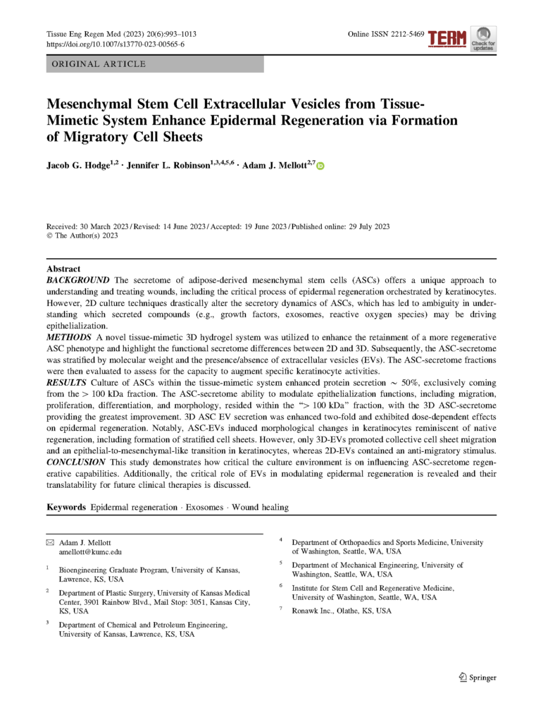
Jacob G. Hodge, Jennifer L. Robinson, Adam J. Mellott
Tissue Eng Regen Med (2023) 20(6):993–1013 Online ISSN 2212-5469
Received: 30 March 2023 / Revised: 14 June 2023 / Accepted: 19 June 2023 / Published online: 29 July 2023
Introduction & Background
Skin is the human body’s largest organ, and it serves as a protective barrier. In response to injury skin begins a well-characterized physiological wound healing process that involves:
-
Migration of cells to injury site
-
Proliferation of appropriate cell type(s)
-
Stimulation of neotissue formation
-
Modulation of cellular biophysical/mechanical structures
One of the key cell types involved in these wound-healing steps is the keratinocyte. These are cells that produce keratin, which is a key protein that forms the outermost layer of the skin. As part of the physiological wound healing process, these cells can increase regenerative capabilities, so they are more able to participate in wound healing. Ultimately, the goal is the re‑epithelialization of the wound site – wound closure protects the wound and deeper tissue structures. For this process to be successful these cells must be able to migrate and proliferate successfully in the right place at the right time (steps 1 and 2 in the list above).
Inadequate keratinocyte activity can result in delayed re-epithelialization, which has higher risks for adverse wound healing outcomes. These can include an overall risk of patient morbidity, development of chronic wounds, required surgical intervention, or even amputation. New research into therapies that can augment keratinocyte behavior could help improve wound healing outcomes and avoid chronic wound development or other negative outcomes.
Mesenchymal stem cells (MSCs) are a promising target for wound healing therapy research. These cells have an innate regenerative capacity and are found in tissues like dermal adipose tissue and hair follicles. This is in part due to the compositional plasticity of their secretory profile. These cells can adjust the type and abundance of secreted products they produce in response to environmental cues, which are abundant in wound healing.
Extracellular vesicles (EVs) are a part of the secretome that may play a key role in wound healing-related signaling. There are a range of EV subtypes which include apoptotic bodies, microvesicles, and exosomes. These three subclasses of EVs can be generally separated as a function of size, and it is unclear what specific size fraction(s) is driving wounding activity like augmenting keratinocytes.
The composition and quality of these EVs are dependent on a range of factors, and they are a reflection of the cell’s environment. In a research setting, this means the EVs produced by cultured stem cells are a reflection of the artificial cell culture environment. Traditionally that has been 2D T-flasks. Cells grow as 2D monolayers in this rigid plastic environment. These unphysiologic conditions can result in stem cell populations that are of variable health and quality. The tissue-mimetic environment of Ronawk’s Bio-Blocks provides an alternative culture environment that more closely mimics in vivo conditions due to the physical properties of the hydrogel block material (manuscript, Table 1). This culture environment produces healthier stem cells that have lower rates of senescence and maintain stemless longer than cells grown in 2D flasks.
This study was designed to explore whether EVs produced by stem cells grown in Bio-Blocks stimulate different wound healing activity in treated cell populations than EVs produced by stem cells grown in 2D flasks.

fig 2. – Mesenchymal Stem Cell Extracellular Vesicles from Tissue-Mimetic System Enhance Epidermal Regeneration via Formation of Migratory Cell Sheets
Complete methods are available in the manuscript PDF. A partial summary is provided below.
Cell Types & Culture
Human, adipose-derived mesenchymal stem cells and human keratinocytes were used for this study. Younger stem cells (donor age: 23 years) and older keratinocytes (donor age: 62 years) were selected to better replicate a potential clinical scenario. Younger keratinocytes perform regenerative wound healing more effectively than older keratinocytes.
2D Culture:
Stem cells and keratinocytes were separately seeded in 2D plastic culture vessels before subculturing for use in reseeding new plastic vessels or experimental assays.
Bio-Block Culture
Bio-Blocks were placed in a glass 6-well plate for cell culture. Stem cells are seeded in a dropwise fashion on the top surface of the block. Following seeding the blocks were submerged in cell culture media. Bio-blocks do not require passaging.
Secretome Isolation, Stratification, & Purification
The media in which cells had been cultured contains the secretome, so to collect the secretome the cell culture media was collected. The media underwent a series of centrifugation filtrations to separate different ranges of molecular weight products (>100 kDa, 100 to 30 kDa, 30 to 10 kDa, and 10 to 3 kDa (manuscript, Figure 2a)). Extracellular vesicles were also isolated from collected conditioned media via centrifugation using a > 100 kDA filter (manuscript, Figure 4a). They were precipitated from this filtered solution using an assay kit. These stratified and purified products can then be used to treat cell culture as needed.
Results & Discussion
Complete results with statistical analysis and a comprehensive discussion of the results are available in the manuscript PDF. A summary is provided below.
Stem cells cultured in the Bio-Blocks displayed enhanced secretion of proteinaceous compounds when compared to the same cells cultured in a 2D environment (manuscript 2B). More protein was made by the Bio-Block cultured stem cells in general in addition to the > 100 kDa fraction being larger as well. Further results revealed that this Bio-Block advantage extended to EV production as well (manuscript, Figure 3A). Stem cells cultured in the Bio-Blocks produced more EVs than cells cultured in a 2D environment.
These secreted products were used to treat populations of keratinocytes to observe changes in behavior. This allowed for data to be collected about the signaling potency of secreted compounds. Keratinocytes treated with EVs produced from within a tissue-mimetic system displayed increases in metabolic activity, proliferation, and migration (manuscript, Figures 4B, C, and D, respectively). These 3 classes of changes are instrumental for successful re-epithelialization of a wound. Extracellular vesicles produced from within the tissue-mimetic culture platform had more potent re-epithelialization stimuli than EVs produced from 2D culture.
To further understand the effects of these EVs, keratinocytes were dosed with one of three concentrations of EVs (low, mid, or high doses) and the effects on 3 unique types of keratin found in the skin. Treatment with the highest dose of EVs produced in a tissue-mimetic environment revealed increases in the expression of all three types of keratin at both the protein and RNA levels (manuscript, Figures 5A and B). This indicates a dose-dependent effect of the extracellular vesicles produced by stem cells in a tissue-mimetic environment.
This study highlights the importance of investigating and understanding the impact of the cell culture environment on stem cells and the products they produce and secrete. This is illustrated by two broad types of specific findings. First, the impact of cell culture environment on the specific secretory activity of stem cells; second, how EVs produced in different culture environments have distinct modulatory effects on keratinocyte activity.
Download The Full Paper
Want to Hear more on our Cell Growth Technology?
Subscribe to get updates on news, case studies, white papers, & more.
Interested in Reading More?


